Original source: Physics World
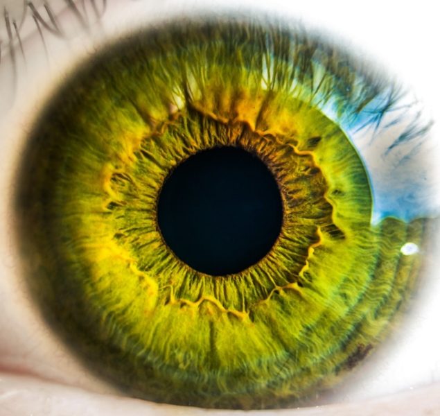
Vision loss is a serious and common symptom of corneal injuries, with more than 1.5 million cases of corneal blindness reported every year. Corneal injuries typically result from a trauma or an infection to the cornea. These can cause scarring and stromal thinning, which can lead to visual impairment and even blindness.
Researchers at the University of California Los Angeles (UCLA), Harvard Medical School and Northeastern University have now developed a biomaterial named GelCORE, which aids the sealing and repair of corneal tissue. The biomaterial is comprised of a gelatin-based adhesive and photoinitiators. Published in Science Advances their research shows how the biomaterial hardens when exposed to visible light, producing a transparent hydrogel with similar biomechanical features to that of the cornea. This adheres to the cornea and encourages regeneration of cells and tissue to repair any damage at that site.
The GelCORE advantage
This technology provides an alternative solution to the current standard treatment options available, which include the use of synthetic glues, corneal transplants and surgery to graft tissue onto the cornea. Both surgery and transplantation risk post-transplant complications such as infection or rejection, and the glues in current use are associated with low biocompatibility, poor transparency, and are undesirably rough and difficult to handle.
Other adhesive materials developed for eye injuries use ultraviolet light, which can introduce toxicity issues. The use of visible blue light to activate the functionalized gelatin adhesive is unique to GelCORE, giving it an important advantage.
Preclinical trial success
The researchers trialled their GelCORE product in preclinical rabbit models of a corneal injury. Here, they made a 3mm long partial cut in the rabbit’s cornea and applied GelCORE concentrations of 20% to the wound, which was then exposed to visible light for 4 minutes.
They found that GelCORE adhered firmly to the damaged tissue in the cornea. Observations over the subsequent day found no inflammation at the site, and a smooth surface across the cornea. After one week of application, they were still able to observe GelCORE at the site on the cornea they had damaged and the product remained transparent. Over a few weeks, the researchers were able to identify the formation of new cell tissue forming, indicating tissue regeneration.
The group additionally showed that by adjusting the concentration and the amount of time exposed to light, the properties of the GelCORE material could be altered. This versatility in the properties of the GelCORE is exciting as it enables product modification and the ability to tailor the product to different patient needs.